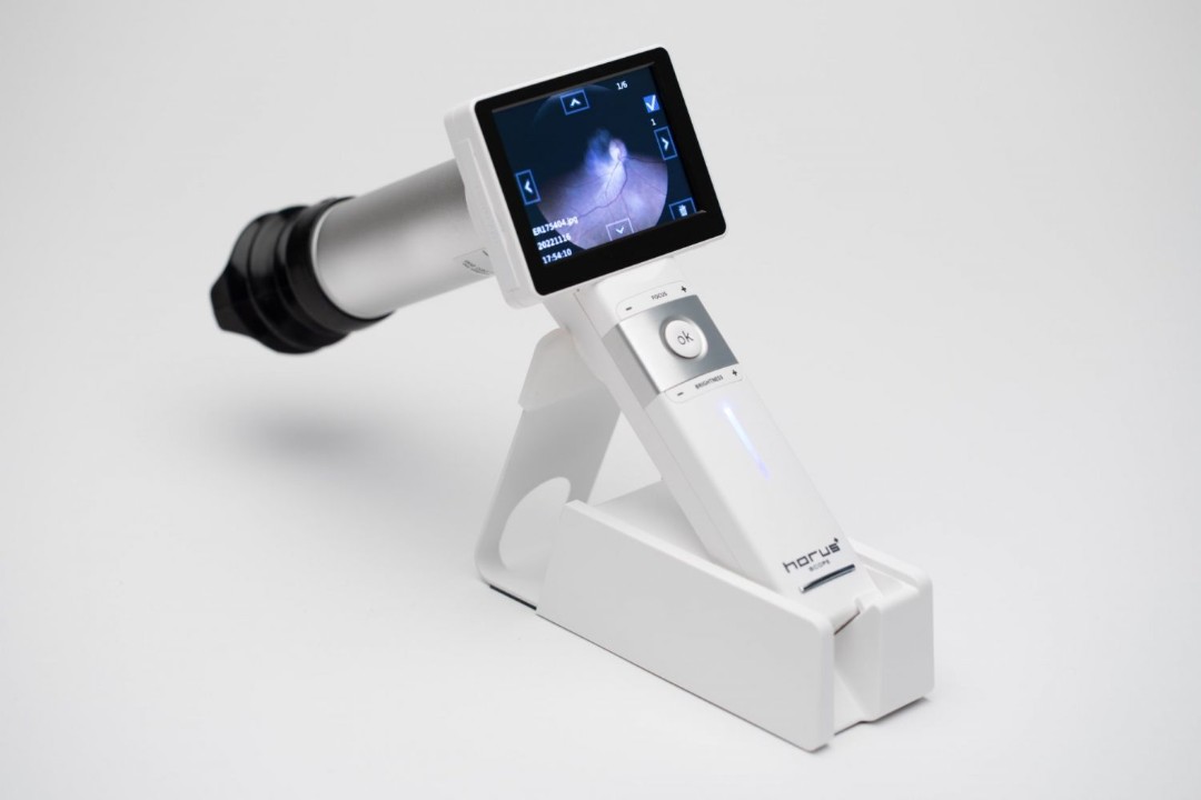
OOphthalmic visualisation using an ophthalmoscope camera system allows the clinician to visualise and capture images of the fundus and optic nerve by using both IR and LED to easily capture images without the need for dilation. Enables non-mydriatic fundus examinations with reflection-free imaging and a wide field of view to capture the appearance of optic disc, macula and retinal vasculature.
| Image Sensor | 2 MPixel CMOS image sensor |
| Dimensions | 89 mm x 44 mm x 205 mm (3.5” x 1.7” x 8”) |
| Weight | 254 g (0.56 lbs) |
| Image format | JPEG |
| Video format | H.264 |
| Display | 3.5” HD full color TFT-LCD display |
| Resolution | full HD true 1920 x 1080 pixels |
| Connector | Bayonet connector for attached optic modules |
