Donor corneal analytics for eye banks and corneal surgeons
Introducing the first multi-imaging system for donor corneal analysis. CellChek D+ provides an amazing view into the cornea that simply has never been seen before defining structure of endothelium, stroma, and epithelium. Comprehensive imaging to assist both eye banks and corneal surgeons to make better decision on implantable material.

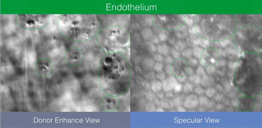
CellChek D+ features a patent applied for “Donor Enhance” imaging system that provides remarkable views of endothelium, intra-stomal structure, and the epithelium. The range of new information from which to make better clinical decisions is spectacular (pun intended): blood cells, fungus, rough keratome cuts, dead cells, epithelial anomalies. Never have we had this view of pre-implanted material. Corneal surgeons are finding this information to be illuminating in helping to understand graft success and failure.
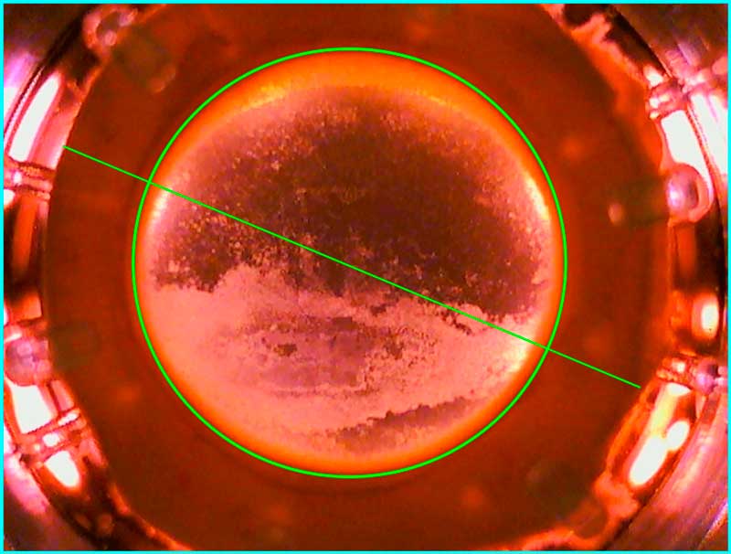
CellChek D and D+ feature a new finder feature with digital measuring / documenting tools.


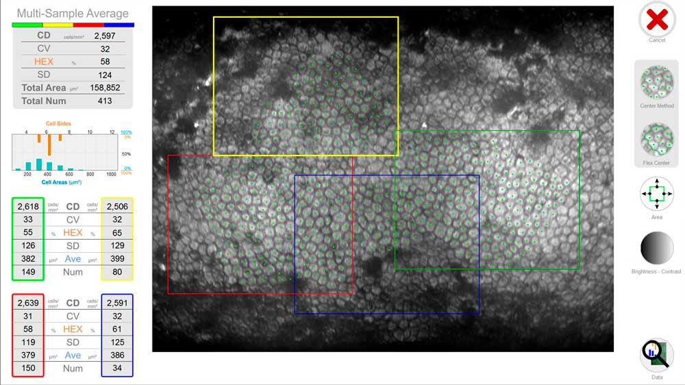
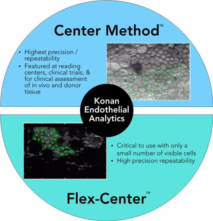
All Konan specular microscopes feature the Center Method of analysis. Center Method is mentioned in FDA panel minutes as being the “gold standard” and is used by virtually every professional reading center for independent assessment of corneal endothelial analytics. Unlike full-auto assessments, the Center Method provides high precision and repeatability for specular images in which relatively small continuous areas of visible cells are visible.
The Flex-Center method is the third tool for advanced stage diseased corneas in which only a very few number of cells are visible. With this semi-automated, perimeter-count method, again, high precision and repeatability is achieved. Only Konan provides the rich set of analytic tools for reliable assessment of the entire spectrum of corneal conditions.
It is not surprising that Konan is the global gold standard in corneal endothelial assessment.
CellChek D and D+ provide a total picture of the cornea. Use digital measurement tools to identify and measure the cornea diameter and defects / scars.

| CellChek D+ | EB-10 | |
|---|---|---|
| Viewing Field | 1000 x 750 µm | 480 X 600 µm |
| Analysis Area (each multi-field area) | 400 X 300 µm X 4 areas (max 480,000 µm2) |
200 X 280 µm |
| Chamber Compatibility | Numedis Life 4C®, Krolman®, B+L®, Stephens® | Life 4C®, Alcon®, Bausch+Lomb®, Krolman® |
| Vial Dimensions | up to 35 mm diameter | up to 35 mm diameter |
| Thermometer Range | 0° to 45° C | 0° to 45° C |
| Stage Translation Ranges | X, Y, and Z = 16 mm, Tilt = 15° | X and Y = 16 mm, Z= 20 mm, Tilt: 5° |
| Illumination | Halogen | LED: primary wavelength 525 nm |
| Cameras | Dual CMOS: Finder and cornea | Dual CMOS: Finder and Cornea |
| Display | Digital data feed to computer | On-board LCD: temperature and pachymetry |
| Electrical | 100-240 VAC, 50/60 Hz, 50 VA | 100-240 VAC, 50/60 Hz, 50 VA |
| Size | 280 (W) X 215 (H) X 265 (D) mm | 200 (W) X 255 (H) X 220 (D) mm |
| Data Interface | USB 2.0 | USB 2.0 |
| Weight | 7.5 kg (without computer) | 6.1 kg (without computer) |
| Operating Conditions | Ambient temp: 10° to 40° C Relative humidity: 30% to 85% Atmospheric pressure: 70 to 106 kPa |
Ambient temp: 10° to 40° C Relative humidity: 30% to 85% Atmospheric pressure: 70 to 106 kPa |
| Regulatory | CE Mark, In Vitro USA | CE Mark, In Vitro USA |
