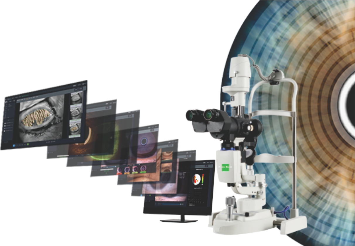
As an excellent dry eye device, our dry eye diagnostic system enhances accurate diagnoses and earlier intervention, providing guidance for customized treatment.
Dry eye diagnosis/Anterior Segment Photography/Lens fitting/Patient management/Telemedicine
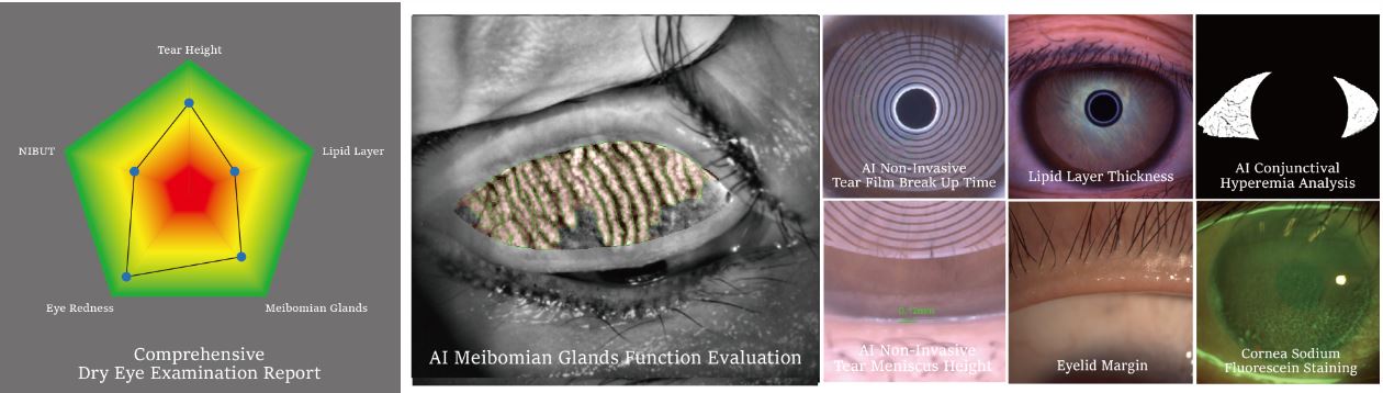
Guided examination:providing a comprehensive report covering 7 dry eye diagnosis.Non-invasive examination,Quantitative data.
Full-automatic Firefly digital module ,easy operation without parameter settings.High quality optics and built-in yellow filter efficiently increase the accuracy of lens fitting.
Professional 1/1.8-inch sensor and 2.4μm pixel,real-time playing and storage.
Smart patient management system,DICOM supported.

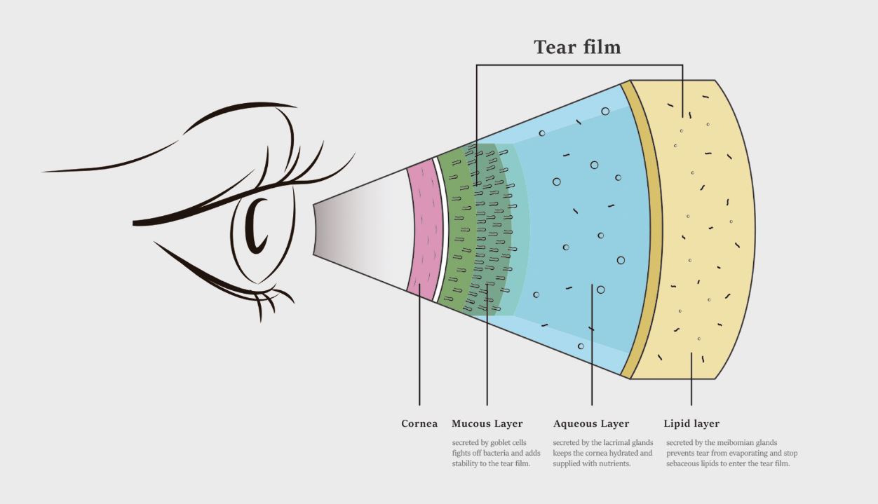
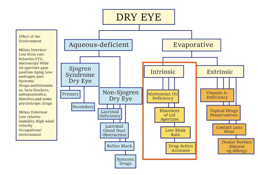
Due to various causes of Dry Eye Disease,traditional examination is difficult to find out the cause and quantify for the diagnosis.
MediWorks Dry Eye Diagnostic System can provide standardized examination and quantified causes evaluation for Dry Eye Disease.
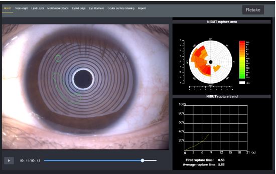
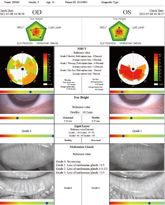
After taking one video, it brings out automatis result of NIBUT and Tear Meniscus Height.
AI identifies the break-up area and analyzes NIBUT automatically.
Fully automatic analysis system provides efficient quantified evaluation for the overall stability of tear film.It automatically acquires the first break up time, average break up time, break up distribution,break up area percentage curve and time distribution.
Grade 0 Normal, First Rupture Time: 10 s Average Rupture Time: 14 s
Grade 1 Warning, First Rupture Time: 6-9 s Average Rupture Time: 7-13 s
Grade 2 Dry eye, First Rupture Time: 5 s Average Rupture Time: 7 s
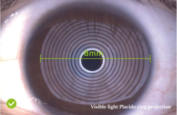
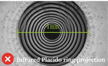
MediWorks adopts Placido ring projection system with visible light to do NIBUT examination,the examination scope is up to 8mm cornea diameter which brings much more comprehensive diagnosis outcome.
The non-invasive examination avoids the irritation brought by the traditional Cornea Sodium Fluorescein Staining.
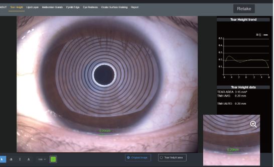
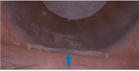
Normal: ≥0.2mm
AI identification system depicts Tear Meniscus area and measures the tear height automatically. Evaluate tear secretion amount and continuity objectively.More efficient and less irritation compared with the traditional Schirmer’s test.

Insufficient tear secretion.
Abnormal dynamics and conjunctival chalasis
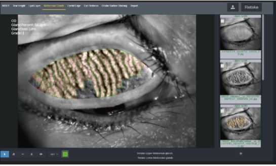
Get original/enhanced/result images by one click AI identification system automatically anlalyzes meibomian glands loss caused by meibomian glands dysfunction with precise and quantified diagnosis results.
Built-in infrared lighting system helps doctors obtain larger image scope of the meibomian glands.
Adjustable depth of field makes the glands more prominent and distinguishable against the background.
Grade 0: No Meibomian Glands Loss
Grade 1: Meibomian Glands Loss < 1/3
Grade 2: Meibomian Glands Loss 1/3-2/3
Grade 3: Meibomian Glands Loss >2/3
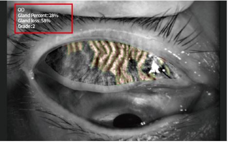
Meibomian glands loss

Image of Meibomian Glands under high-magnification
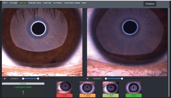
White ring projection system ensures a larger examination area compared to Placido ring.By comparing with the standard grading template and recording the Lipid Layer thickness, it is helpful for judging MGD.
Grade 1: <30
Grade 2: 30-60
Grade 3: 60-80
Grade 4: >80
(Unit:mm)
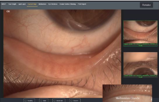
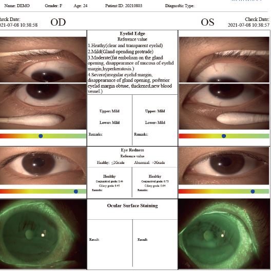
MediWorks professional design of optical system is capable of providing HD digital image that remains clear and sharp even zoom in, meets the examination requirements of the overall shape of eyelid margin and its slight change.
2. Mild including (gland cap crown - glandular prominent)
3. Moderate including (glandular fat plug - disappearance of the marginal mucosa, hyperkeratosis)
4. Severe including (uneven margins, disappearance of the meibomian glands - posterior margin Blunt round, thickening, new blood)
Normal: ≤2 Abnormal: >2
The unique AI identification system can identify and calculate percentages of conjunctival congestion and ciliary congestions and evaluate severity of eye congestion.


Effectively increases positive rate of early corneal epithelial staining.
Built-in yellow filter along with cobalt-blue filter makes the corneal sodium fluorescein images more clearly.
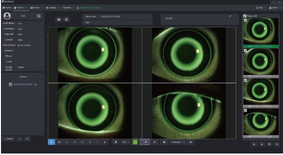
Comparison of Patient records Smart Patient Management system supports repeated comparison among medical records to help doctors develop customized treatment plans and evaluate treatments.
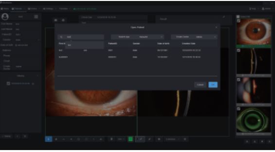
Patient Management system allows doctors to build and edit medical records, and quickly search the patient case by key words. Besides, doctors can note patients’ situation via the software. With the DICOM-supported system, Mediview is connected with medical systems in hospitals.