As part of the training process Mercoframes offers to customers and distributors, today we speak about: Welch Allyn 3.5 V AutoStep® Coaxial Ophthalmoscope Head, 11730 Thank you for follow us. To order: sales@mercoframes.com 1-312-962-7227 Alex Gutman |CEO Mercoframes
Coaxial optics produce a shadow-free spot, easier entry into undilated pupils, and a larger field of view 18 aperture/filter combinations for greater versatility: micro, small, and large spot sizes, cobalt blue filter for corneal exams, fixation target, or slit aperture can be combined with red-free filter, polarizing filter, or unfiltered Halogen HPX illumination 68 lenses in single-diopter steps for precise resolution (+38 to -30)
Sealed optics keep out dust and dirt for years of maintenance-free operation
Rubber brow rest prevents scratching of eyeglasses
Transparency of the cornea, lens and vitreous humor permits the physician to directly view arteries, veins, optic nerve and the retina. Direct observation of the structures of the fundus through an ophthalmoscope may show disease of the eye itself or may reveal abnormalities indicative of disease elsewhere in the body. Among the most important of these are vascular changes due to diabetes or hypertension and swelling of the optic nerve head due to papilledema or optic neuritis. In this sense, the eye serves as a window through which many valuable clinical evaluations may be made. When a preliminary diagnosis of an imminently dangerous eye condition, such as acute glaucoma or retinal detachment, is made by the examiner, prompt referral to an ophthalmologist may prevent irreversible damage. Or, when distressing but less urgent conditions, such as visual impairment due to cataract or vitreous floater are recognized, the patient can be reassured and referred.
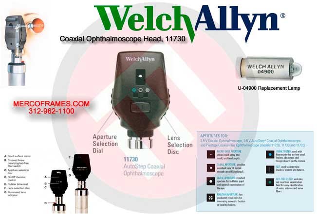
Standard apertures
There is a wide range of practical apertures to select from: micro spot, small spot, large spot, fixation, slit and cobalt blue filter. A red-free filter is also available for use on the apertures. This selection of apertures covers all the physician’s needs in an ophthalmologic examination.
Other filters
Welch Allyn ophthalmoscopes #11720 and #11730 are equipped with a unique sliding switch that greatly increases their versatility.
Red-free filter: When the switch is positioned to the left (while facing the instrument front) it will be beneath a green dot and the red-free filter will be in place. This can be used in conjunction with any aperture. The red-free filter excludes red rays from the examination field; this is superior to ordinary light in viewing slight alterations in vessels, minute retinal hemorrhages, ill-defined exudates and obscure changes in the macula. The nerve fibers become visible and the observer may note the disappearance of such fibers, as in optic nerve atrophy. The background appears gray, the disc appears white, the macula appears yellow, the fundus reflex is intense and the vessels appear almost black. This filter is also used to help distinguish veins from arteries...veins stay relatively blue, but oxygenated arterial blood makes arteries appear blacker. This makes differentiation easier for the examiner.
Crossed linear polarizing filter: When the switch is positioned to the right (while facing the instrument front) it will be beneath a while circle with crosshairs inside. The crossed linear polarizing filter will be in place. This filter is used to eliminate corneal glare and reflection and can be used with any aperture.
Additional uses for the ophthalmoscope
In addition to examination of the fundus, the ophthalmoscope is a useful diagnostic aid in studying other ocular structures. The light beam can be used to illuminate the cornea and the iris for detecting foreign bodies in the cornea and irregularities of the pupil. By placing the +15.00 lens in the scope and looking at the pupil as in fundus examination [2 inches (5cm) distance from patient], the physician may verify doubtful pupillary action.
The practitioner can also easily detect lens opacities by looking at the pupil through the +6 lens setting at a distance of 6 inches (15cm) from the patient. In the same manner, vitreous opacities can be detected by having the patient look up and down, to the right and to the left. Any vitreous opacities will be seen moving across the pupillary area as the eye changes position or comes back to the primary position.
How to Conduct an Ophthalmologic Examination
Position the ophthalmoscope about 6 inches (15cm) in front and 25° to the right side of the patient. (Step 5)
In order to conduct a successful examination of the fundus, the examining room should be either semi-darkened or completely darkened. It is preferable to dilate the pupil when there is no pathologic contraindication, but much information can be obtained through the undilated pupil.
The following steps will help the physician obtain satisfactory results:
CAUTION:
Before actuating the red-free filter/crossed linear polarizing filter slide switch, pull the instrument away from the patient’s face to prevent contact with finger or switch.
Examine the disc for clarity of outline, color, elevation and condition of the vessels. (step 7)
Overcoming corneal reflection One of the most troublesome barriers to a good view of the retina is the light reflected back into the examiner’s eye by the patient’s cornea — a condition know as corneal reflection.